Endoscopes transmit LED light sources via fibre optic bundles and incorporate cameras for imaging, representing the typical operational mode of modern endoscopic technology. This design is widely applied in fields such as medical diagnosis and industrial inspection. Below, we break down the technology from three key angles: technical principles, core components, and application scenarios.
1. Overview of Technical Principles
The core function of an endoscope is to achieve illumination and imaging within enclosed or narrow spaces. Its operational process can be divided into three steps:
- Light Source Transmission: Visible light (or specific wavelength light, such as ultraviolet light for fluorescence imaging) emitted by the LED is transmitted through the fibre optic bundle to the front end, illuminating the observation area.
- Image Acquisition: The built-in miniature camera (typically a CMOS or CCD sensor) captures the optical signals from the illuminated area and converts them into electrical signals.
- Signal Processing and Display: The electrical signals are transmitted via cables to external devices (such as displays or computers), processed, and presented as clear images.
2. Core Components and Their Functions
LED Light Source
- Advantages: Compact size, low power consumption, long lifespan, capable of providing stable white light or specific wavelengths of light (such as near-infrared light for vascular imaging).
- Control: Brightness is adjusted via external circuits to adapt to the reflective properties of different tissues or objects.
Fibre Optic Bundle
- Structure: Composed of thousands of extremely thin glass or plastic fibres, each fibre transmits the optical signal for a single pixel.
- Function: Can transmit illumination light (“illumination fibre optic bundle”), and some endoscopes also transmit images via another set of fibre optic bundles (“imaging fibre optic bundle,” suitable for traditional models without built-in cameras). However, in modern mainstream designs, fibre optic bundles primarily handle illumination, while imaging is performed by the camera.
Built-in Camera
- Miniaturised design: Sensor size typically ranges from a few millimetres (e.g., 1/10-inch CMOS), paired with a miniature lens assembly, ensuring the endoscope probe remains slender (diameter as small as 2–3 millimetres, suitable for minimally invasive surgery).
- Resolution: Medical endoscope cameras typically have resolutions of 1080p or higher, with some high-end models reaching 4K, to meet detailed observation requirements (e.g., identifying early-stage lesions).
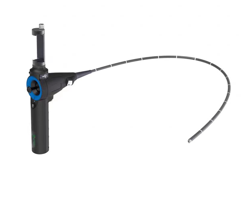
Probe and Housing
- The probe’s front end integrates an illumination window and camera lens, while the housing is made of biocompatible materials (e.g., medical-grade stainless steel or polymer materials) to ensure safe insertion into bodily cavities.
- Some models feature adjustable bending functionality (controlled via a rear knob to manipulate the steel wire), facilitating access to complex structures (e.g., gastrointestinal tract, respiratory tract).
3. Application Scenarios
Medical Field
- Diagnosis: Gastroscopes, colonoscopes, laryngoscopes, etc., capture images of the inner walls of the digestive and respiratory tracts to assist in detecting inflammation, polyps, tumours, and other lesions.
- Surgery: Laparoscopes, arthroscopes, etc., provide real-time visualisation of internal structures during minimally invasive surgeries, guiding doctors in procedures such as lesion removal and tissue suturing.
Industrial Inspection
Used for non-destructive testing of enclosed structures such as pipelines, engine blocks, and circuit boards, imaging is employed to identify defects such as cracks, corrosion, or foreign objects.
Other Fields
- Archaeology: Inspecting the internal structures of cultural relics (e.g., layers beneath murals).
- Security: Detecting abnormal conditions in confined spaces (e.g., inside walls, gaps in equipment).
Technical Advantages
- Minimally invasive: Compared to traditional surgery, the combination of fibre optic transmission and miniature cameras significantly reduces the diameter of the endoscope, minimising trauma.
- Real-time: Low image transmission latency (typically < 100 milliseconds) meets real-time observation requirements.
- Flexibility: The fibre optic bundle is bendable and, combined with an adjustable probe, can adapt to complex space detection.
This design highly integrates lighting and imaging functions, serving as the core technological foundation for endoscopes to achieve “visualised detection.” It continues to evolve towards higher resolution, smaller size, and greater intelligence (e.g., AI-assisted lesion identification).
The Arrangement of Fibres in Fibre Optic Bundles
The primary function of the illumination fibre bundle in an endoscope is to transmit light from the light source, providing illumination for the observation area. The individual fibre strands have relatively thick diameters, typically 15–30 micrometres, and their arrangement does not require uniformity, belonging to a non-coordinated arrangement. This means that the individual fibres in the illumination fibre bundle do not have a one-to-one correspondence between their input and output ends. Common arrangement forms include circular, linear, rectangular, hexagonal, ring-shaped, mixed circular, and crescent-shaped.
We provide tailored solutions for fibre optic bundles in endoscopes—contact us to learn more!